
de Female Human Anatomy Organs Diagram mar webmds abdomen anatomy page
Breasts. Summary. Female anatomy includes the external genitals, or the vulva, and the internal reproductive organs, which include the ovaries and the uterus. One major difference between males.

Female Anatomy Upper Body Stock Photo Download Image Now iStock
These internal structures of female anatomy include the: Vagina: The vagina is a muscular canal that connects the cervix and the uterus. It leads to the outside of the body. Parts of the vagina are made of collagen and elastin, which help it expand during sexual stimulation and childbirth. Cervix: The cervix is the lower part of the uterus that.

Female Pelvis and Abdomen Comparison TrialExhibits Inc.
Female pelvis bones. Hip bones. There are two hip bones, one on the left side of the body and the other on the right. Together, they form the part of the pelvis called the pelvic girdle.

Human Anatomy Abdomen Anatomy Pinterest
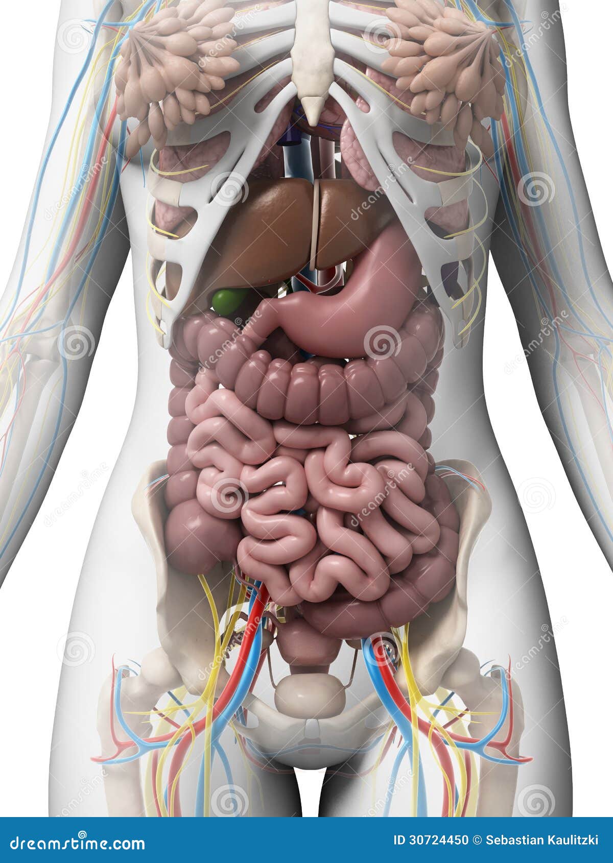
Stomach. The stomach is on the upper-left area of the abdomen below the liver and next to the spleen. It stores and breaks down the foods and liquids we eat before they move to digestion. When the.
Anatomy Of The Female Abdomen And Pelvis, Cut away View
3. Anterior view: anatomy of female abdomen and pelvis: stomach and omentum. 4. Anterior view: anatomy of female abdomen and pelvis: small bowel and colon. 5. Anterior view: anatomy of female abdomen and pelvis: peritoneum. 6. Anterior view: anatomy of female abdomen and pelvis: posterior wall of abdomen. BOARD 2.
Human Abdomen Anatomy Female Female Colon With Abdominal Organs
Browse 617 female anatomy diagram photos and images available, or start a new search to explore more photos and images. Browse Getty Images' premium collection of high-quality, authentic Female Anatomy Diagram stock photos, royalty-free images, and pictures. Female Anatomy Diagram stock photos are available in a variety of sizes and formats to.

Abdomen Wikipedia, la enciclopedia libre
Browse 1,154 human body organs anatomy in women photos and images available, or start a new search to explore more photos and images. Browse Getty Images' premium collection of high-quality, authentic Human Body Organs Anatomy In Women stock photos, royalty-free images, and pictures.

Anatomy of a Female Abdomen TrialExhibits Inc.
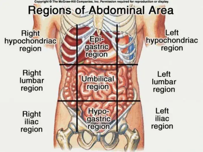
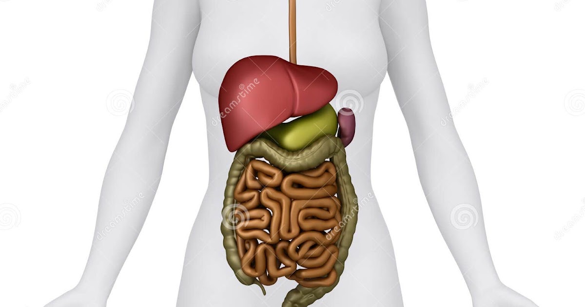
Pregnancy Symptoms Week by Week ». This Article is the detailed account of all the major organs that are categorized under the nine regions in the abdominal cavity 1) Stomach 2) Intestines a) Small Intestine Duodenum Jejunum Ileum b) Large Intestine Ceacum Colon (Ascending, Transverse and Descending) Rectum Anal Canal 3) Liver 4) Gall bladder.
Abdomen AnatomyFemale Female Abdominal Anatomy Illustration Stock
The abdominal wall surrounds the abdominal cavity, providing it with flexible coverage and protecting the internal organs from damage. It is bounded superiorly by the xiphoid process and costal margins, posteriorly by the vertebral column and inferiorly by the pelvic bones and inguinal ligament.. The abdominal wall can be divided into two sections: anterolateral and posterior abdominal walls.
Abdominal Anatomy Pictures Female Female abdominal anatomy, computer
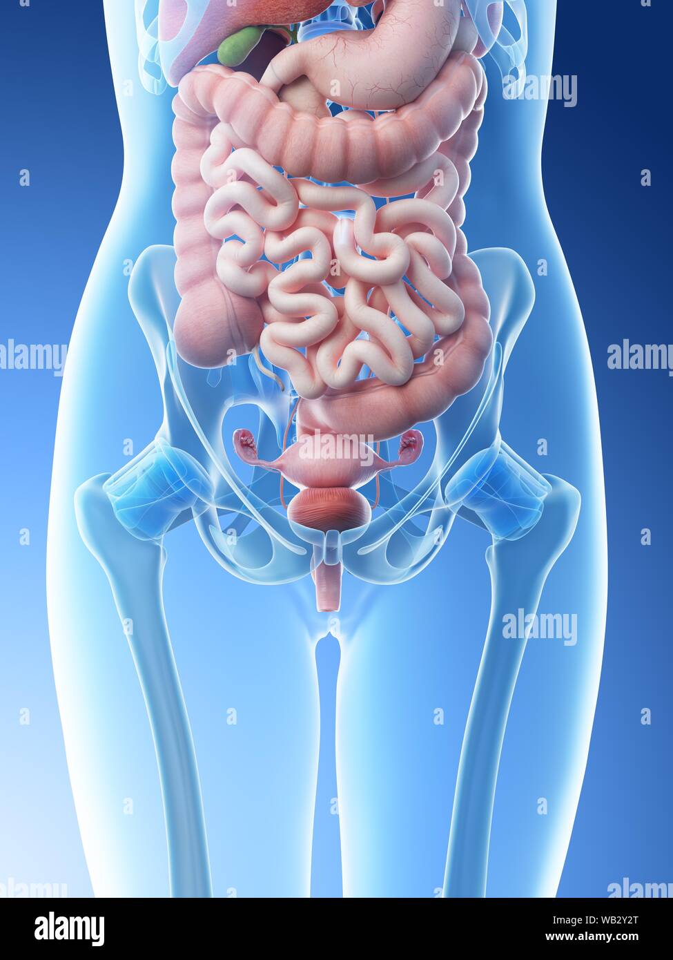
The abdomen and pelvic regions are continuous with each other, making up the distal part of the trunk. Bar the brain, heart and lungs, this region contains virtually all your body organs, including those involved in the digestive, endocrine, lymphatic, urinary and reproductive systems. So, it is crucial that you cover this section thoroughly.
Abdominal Anatomy Pictures Female Abdominal Viscera Posterior Human
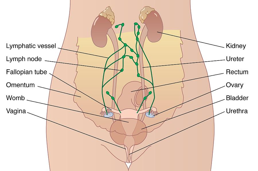
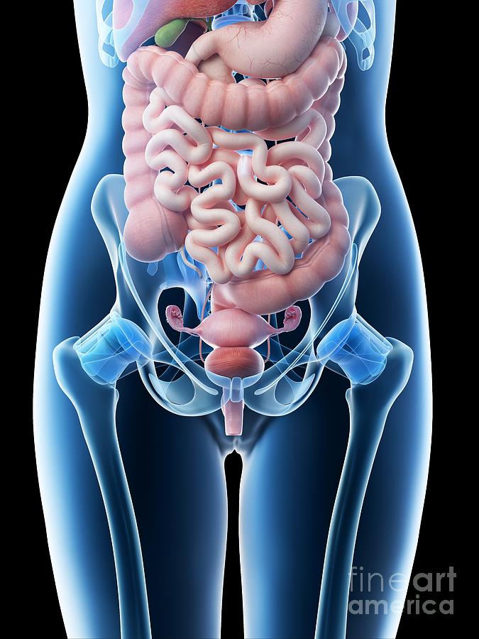
Anatomy of Female Pelvic Area. Endometrium. The lining of the uterus. Uterus. Also called the womb, the uterus is a hollow, pear-shaped organ located in a woman's lower abdomen, between the bladder and the rectum. Ovaries. Two female reproductive organs located in the pelvis. Fallopian tubes.

Human Anatomy Female Abdomen Female Anatomy Of Abdomen Anatomy
The muscles of the abdomen protect vital organs underneath and provide structure for the spine. These muscles help the body bend at the waist. The major muscles of the abdomen include the rectus.

Abdominal Wall Muscle Anatomy Abdomen Muscle Anatomy Muscle anatomy
ID: exh6130a. This medical illustration depicts a mid-sagittal view of the normal anatomy of the female abdomen and pelvis. Labeled structures include the large bowel (colon or large intestine), umbilicus, small intestine, ovary, fallopian tube, uterus and bladder.

Female Abdomen Anatomy Quadrants / Abdominal Surface Anatomy Radiology
Picture of Abdomen. The abdominal cavity is the part of the body that houses the stomach, liver, pancreas, kidneys, gallbladder, spleen, and the large and small intestines. The diaphragm marks the top of the abdomen and the horizontal line at the level of the top of the pelvis marks the bottom. Connective tissue called the mesentery holds the.
Female anatomy stock illustration. Image of hepatic, medicine 30724450
The female reproductive system is an intricate arrangement of structures that can separate into external and internal genitalia. The external genitalia comprises the structures outside of the true pelvis, including the labia majora and minora, vestibule, Bartholin glands, Skene glands, clitoris, mons pubis, perineum, urethral meatus, and periurethral area. The internal genitalia is the.

Aliviar Mezquita Arne abdominal anatomy diagram curva Nuevo significado
The vulva is the global term that describes all of the structures that make the female external genitalia. The components of the vulva are the mons pubis, labia majora, labia minora, clitoris, vestibular bulbs, vulva vestibule, Bartholin's glands, Skene's glands, urethra, and vaginal opening. The mons pubis is a tissue mound made up of fat.